Common Types of Heart Defects

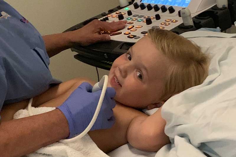
Jackson was born with tricuspid atresia, hypoplastic right ventricle, atrial septal defect and a ventricular septal defect.
Congenital heart defects are structural problems arising from abnormal formation of the heart or major blood vessels. At least 18 distinct types of congenital heart defects are recognized, with many additional anatomic variations. Ongoing progress in diagnosis and treatment (surgery and heart catheterization) makes it possible to treat most defects, even those once thought to be hopeless.
The Outlook
If your child is born with a heart defect today, the chances are better than ever that the problem can be overcome and that a fulfilling adult life will follow. As diagnosis and treatment continue to advance, treatments other defects will be developed. Your cardiologist will discuss your particular heart defect, treatment options and expected results.
The descriptions and pictures of common heart defects that follow will help you understand the heart problem you or your child are facing. For more in-depth information, use the links which will provide a deeper of explanation of the science and will also answer some common questions such as treatment options, ongoing care needs, and expected limitations or activity levels.
Congenital Defects - A simplified glossary
Healthy Heart Function
A normal heart has valves, arteries and chambers that carry the blood in a circulatory pattern: body–heart–lungs–heart–body. When all chambers and valves work correctly, the blood is pumped through the heart, to the lungs for oxygen, back the heart and out to the body for delivery of oxygen. When valves, chambers, arteries and veins are malformed, this circulation pattern can be impaired. Congenital heart defects are malformations that are present at birth. They may or may not have a disruptive effect on a person's circulatory system.
Learn how the healthy heart works.
Aortic Valve Stenosis (AVS)
A valve from the heart to the body that does not properly open and close and may also leak blood. When the blood flowing out from the heart is trapped by a poorly working valve, pressure may build up inside the heart and cause damage.
More information about Aortic Valve Stenosis.
Atrial Septal Defect (ASD)
A "hole" in the wall that separates the top two chambers of the heart.
This defect allows oxygen-rich blood to leak into the oxygen-poor blood chambers in the heart. ASD is a defect in the septum between the heart's two upper chambers (atria). The septum is a wall that separates the heart's left and right sides.
More information about Atrial Septal Defect.
Coarctation of the Aorta (CoA)
A narrowing of the major artery (the aorta) that carries blood to the body.
This narrowing affects blood flow where the arteries branch out to carry blood along separate vessels to the upper and lower parts of the body. CoA can cause high blood pressure or heart damage.
More information about Coarctation of the Aorta (CoA).
Complete Atrioventricular Canal defect (CAVC)
A large hole in center of the heart affecting all four chambers where they would normally be divided. When a heart is properly divided, the oxygen-rich blood from the lungs does not mix with the oxygen-poor blood from the body. A CAVC allows blood to mix and the chambers and valves to not properly route the blood to each station of circulation.
More information about Complete Atrioventricular Canal defect (CAVC).
d-Transposition of the great arteries
A heart in which the two main arteries carrying blood away from the heart are reversed.
A normal blood pattern carries blood in a cycle: body-heart-lungs-heart-body.
When a d-transposition occurs, the blood pathway is impaired because the two arteries are connecting to the wrong chambers in the heart.
This means that the blood flow cycle is stuck in either:
- body–heart –body (without being routed to the lungs for oxygen) or
- lungs–heart–lungs (without delivering oxygen to the body)
Without surgery, the only way to survive this condition temporarily is to have leakages that allow some oxygen-rich blood to cross into the oxygen-poor blood for delivery to the body. A hospital facility can also catheterize a patient until corrective surgery can be performed.
More information about d-Transposition of the great arteries.
Ebstein's Anomaly
A malformed heart valve that does not properly close to keep the blood flow moving in the right direction. Blood may leak back from the lower to upper chambers on the right side of the heart. This syndrome also is commonly seen with ASD (or a hole in the wall dividing the two upper chambers of the heart).
More information about Ebstein's Anomaly.
I-Transposition of the great arteries
A heart in which the lower section is fully reversed.
This malformation of the heart causes a reversal in the normal blood flow pattern because the right and left lower chambers of the heart are reversed. The I-transposition, however, is less dangerous than a d-transposition because the great arteries are also reversed. This "double reversal" allows the body to still receive oxygen-rich blood and the lungs to still receive the oxygen-poor blood.
More information about I-Transposition of the great arteries.
Patent Ductus Arteriosis (PDA)
An unclosed hole in the aorta.
Before a baby is born, the fetus's blood does not need to go to the lungs to get oxygenated. The ductus arteriosis is a hole that allows the blood to skip the circulation to the lungs. However, when the baby is born, the blood must receive oxygen in the lungs and this hole is supposed to close. If the ductus arteriosis is still open (or patent) the blood may skip this necessary step of circulation. The open hole is called the patent ductus arteriosis.
More information about Patent Ductus Arteriosis (PDA)
Pulmonary Valve Stenosis
A thickened or fused heart valve that does not fully open. The pulmonary valve allows blood to flow out of the heart, into the pulmonary artery and then to the lungs.
More information about Pulmonary Valve Stenosis.
Single Ventricle Defects
Rare disorders affecting one lower chamber of the heart. The chamber may be smaller, underdeveloped, or missing a valve.
Hypoplastic Left Heart Syndrome (HLHS) — An underdeveloped left side of the heart. The aorta and left ventricle are too small and the holes in the artery and septum did not properly mature and close.
Pulmonary Atresia/Intact Ventricular Septum — The pulmonary valve does not exist, and the only blood receiving oxygen is the blood that is diverted to the lungs through openings that normally close during development.
Tricuspid Atresia — There is no tricuspid valve in the heart so blood cannot flow from the body into the heart in the normal way. The blood is not being properly refilled with oxygen it does not complete the normal cycle of body–heart –lungs–heart –body.
More information about Single Ventricle Defects.
Tetralogy of Fallot
A heart defect that features four problems.
They are:
- a hole between the lower chambers of the heart
- an obstruction from the heart to the lungs
- The aorta (blood vessel) lies over over the hole in the lower chambers
- The muscle surrounding the lower right chamber becomes overly thickened
More information about Tetralogy of Fallot.
Total Anomalous Pulmonary Venous Connection (TAPVC)
A defect in the veins leading from the lungs to the heart.
In TAPVC, the blood does not take the normal route from the lungs to the heart and out to the body. Instead, the veins from the lungs attach to the heart in abnormal positions and this problem means that oxygenated blood enters or leaks into the wrong chamber.
More information about Total Anomalous Pulmonary Venous Connection (TAPVC).
Truncus Arteriosus
When a person has one large artery instead of two separate ones to carry blood to the lungs and body.
In a normal heart, the blood follow this cycle: body-heart-lungs-heart-body. When a person has a truncus arteriosus, the blood leaving the heart does not follow this path. It has only one vessel, instead of two separate ones for the lungs and body. With only one artery, there is no specific path to the lungs for oxygen before returning to the heart to deliver oxygen to the body.
More information about Truncus Arteriosus.
Ventricular Septal Defect (VSD)
VSD is a hole in the wall separating the two lower chambers of the heart.
In normal development, the wall between the chambers closes before the fetus is born, so that by birth, oxygen-rich blood is kept from mixing with the oxygen-poor blood. When the hole does not close, it may cause higher pressure in the heart or reduced oxygen to the body.
More information about Ventricular Septal Defect (VSD).


